|
FROM PERIODONTOLOGY
TO PROSTHESIS
|
|
Published in June 1987
|
|
COMPLETE ORAL REHABILITATION
|
|
|
|
|
|
|
|
|
|
|
|
|
|
GUSTAVO
PETTI
Physician and Surgeon specializing in Dentistry. Periodontist.
Piazza Repubblica 4, 09129 Cagliari, Italy.
tel ++39 070 498159, fax ++39 070 400164
web site www.gustavopetti.it
|
|
|
|
|
|
|
|
|
|
|
|
|
|
OBJECTIVE
Exploitation of 4.4 and 4.5 for prosthesis |
|
|
|
|
|
|
|
|
|
|
|
|
|
|
|
|
|
|
|
 |
|
 |
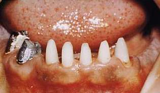
| Fig.
16 The clinical crowns of the two premolars are exposed and easy to work
on. |
Fig.
17 Following construction of the cast gold core and suitable preparation
of the shoulder of both premolars coronally to the gingival attachment,
prosthesic preparation of the all incisors is completed. |
|
|
|
|
|
|
|
|
|
|
|
|
|
 |
 |
|
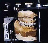
Fig.
18 Transfer of impressions onto the articulator and subsequent proper construction
of the p.g.c. prosthesis by the Brau-Mureddu laboratory of dental mechanics). |
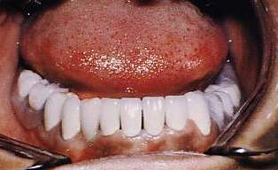
Fig.
19 Porcelain gold-platinum lower prosthesis with framework and precision
attachments with attachments |
|
|
|
|
|
|
|
|
|
|
|
|
 |
|
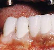
Fig.
20 Detail of the appearance of gum at 4.5 and 4.6 after 26 months. |
|
|
|
|
|
|
|
|
|
|
|
|
|
|
|
|
|
|
|
|
 |
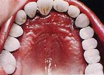 |
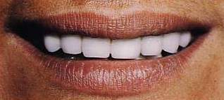
| Figs.
21-22 Upper fixed gold-platinum-platinum prosthesis. |
|
|
|
|
|
|
|
|
|
|
|
|
|
OBJECTIVE
Follow-up to verify the effectiveness of the treatment.
|
|
|
|
|
|
|
|
 |
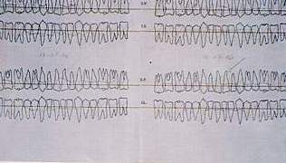 |
| Fig.
23 Situation of bone on graph paper: on the left in May 1984 and on the
right in 1986. |
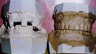
Fig.
24 Patterns of patient's situation before and after rehabilitation: the
problems of vertical dimension, gnathology and prosthesis which have been
eliminated. |
|
|
|
|
|
|
|
|
|
|
|
|
 |
 |
| Fig.
25 Orthopantomography at the beginning of treatment. |
|
|
|
|
|
|
|
|
|
|
|
|
|
|
|
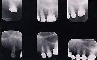
Fig.
26 X-ray of upper incisors at the time of treatment, of endodontal and prosthesic
treatment of 2.2, of the bone implant and finally of the result after 26
months from the beginning of treatment respectively. |
|
|
 |
|
|
|
|
|
|
|
|
|
Fig.
27 Top left and bottom left: the situation of 1.7 and 1.3 respectively;
from left to right and from top to bottom, the situation before the operation,
after 6 months, 12 months and 26 months from the beginning of treatment. |
|
|
|
|
|
|
|
|
|
|
|
|
|
|
|
|
|
|
|
|
|
|
|
 |
|
|
|
|
|
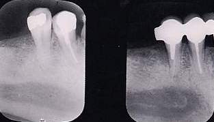
Fig.
28 X-rays before and 13 months after osteoplastics and apical repositioning
of the flap. |
|
|
|
|
|
|
|
|
|
|
|
|
|
|
|
|
|
|
|
 |
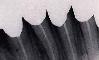 |
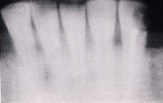
| Figs
.29-30 X-rays of the bone profile of the lower incisors before and 13 months
after bone remodelling. |
|
|
|
|
|
|
|
|
|
|
|
|
|
|
|
|
 |
|
|
|
|
 |
|
 |
 |
|
 |
 |
|
 |
 |
 |
 |
 |
 |