|
Published in November 1989
|
|
|
|
PERIODONTOLOGY
|
|
|
|
|
|
|
|
|
|
|
|
|
|
GUIDED
PERIODONTAL REGENERATION WITH AN AMNIOTIC MEMBRANE AND FIBRIN GLUE
|
|
GUSTAVO
PETTI
Physician and Surgeon specializing in Dentistry. Periodontist.
Piazza Repubblica 4, 09129 Cagliari, Italy.
tel ++39 070 498159, fax ++39 070 400164
web site www.gustavopetti.it
University
of Sassari [Italy]
Post-graduate School of Dentistry and Dental Prosthesis, Prof. V. Tenti,
Director.
Course in Periodontology
held by Prof. G. Petti
|
PRELIMINARY
HISTOLOGICAL AND EXPERIMENTAL CONSIDERATIONS
|
Key
words:
Fibrin glue, Interpore 200, amniotic
membrane, new attachment
The most ambitious
goal in bone surgery, the creation of a new attachment, calls for suitable
materials and special techniques. Only a rigorous in vivo experimental
protocol and subsequent histological examinations can ensure success.
|
|
|
 |
|
|
|
|
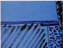
Fig.
1 Cross section, from bottom to top: bone graft (blue), Tissucol layer (yellow),
amniotic membrane (light blue) mucogingival flap (purple). The amniotic
membrane inhibits apical migration of the epithelial attachment. |
Fig.
2 Graft of amniotic membrane B with fibrin glue A as a protection of the
non-re-absorbable hydroxyapatite implant C. Direct contact between bone
and root may lead to root re-absorption and anchylosis. |
|
|
|
|
|
|
|
|
|
|
|
|
|
Basics
of in vivo experimentation
|
|
|
|
|
|
Fig.
3 After preparing the receiving bed for the bone graft, the amniotic membrane
B is glued to the root with fibrin glue A. |
Fig.
4 At this point, after gluing the amniotic membrane, the operation continues
with the Interpore 200 bone implant (C)
|
|
|
|
|
|
|
|
|
|
|
|
|
|
 |
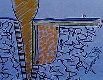
Fig.
5 The implant is covered with the amniotic membrane B saturated with Tissucol
(A') and the mucoperiosteal flap D is repositioned. Periodontal space E
is protected by the amniotic membrane B placed between the bone implant
C and the root, to which the membrane is attached with Tissucol (A'), which
also has the function of biostimulating cells in the periodontal space.
The amniotic membrane B' glued with fibrin glue A' onto the implant C and
well fitted to the root inhibits apical migration of attachment D.
A = fibrin glue
B = amniotic membrane
C = Interpore 200 implant
D = mucoperiosteal flap
E = periodontal space with ligament
A' = fibrin glue
B' = amniotic membrane
|
|
|
|
|
|
|
|
|
|
|
|
|
|
|
|
|
|
|
|
|
|
|
|
|
Discussion
|
| By inhibiting
direct contact between bone and root with the amniotic membrane and fibrin
glue, reabsorption and consequent anchylosis is avoided. By avoiding contact
between the periodontal space and gum, apical migration of the epithelial
attachment is inhibited and, in the long run, this should lead to the formation
of a short epithelial attachment. The amniotic membrane is then glued to
the root In reality, the glue occupies the space between the root and the
membrane and is thus invaded by cells which are capable of regenerating
the periodontal ligament; we must not forget the biostimulating action and
tissue regeneration performed by the fibrin glue and the trophoblastic inner
surface of the amniotic membrane. Between the inner trophoblastic surface
of the membrane (facing the root) and the outer epithelial surface (facing
the bone graft) we find the basal membrane; this sandwich of tissues, especially
owing to the presence of the basal membrane, represents a good barrier against
tissue cell penetration. |
|
|
|
|
|
|
|
|
|
|
|
|
 |
|
|
|
|
|
|
|
|
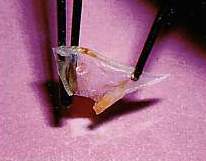
| Fig.
6 Amniotic membrane: the edges of the epithelial face curl inwards; this
is important in distinguishing the epithelial surface from the trophblastic
surface. |
|
|
|
|
|
|
|
|
|
|
|
|
|
|
|
|
|
|
|
|
|
Materials
and methods
|
|
|
|
|
|
|
|
|
|
|
|
|
|
The
amniotic membrane |
|
|
|
|
|
|
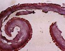
Fig.
7 Cross section of the amniotic membrane (Amniex Mastelli s.r.l.). Note
the curling edges. |
 |
|
|
|
|
|
|
|
|
|
|
|
|
|
|
|
|
|
|
|
|
| Interpore
200 |
|
|
|
|
|
|
|
|
|
|
|
|
|
|
|
|
|
|
|
|
|
|
 |
|
 |
 |
 |
|
|
|
|
|
|
|
|
|