 |
 |
||||||||||||||||
|
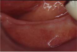
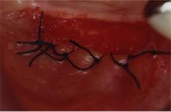
Figs 12, 13.
Mucogingival stripping at the rear right edentate col to increase the fornix. |
|||||||||||||||||
 |
|||||||||||||||||
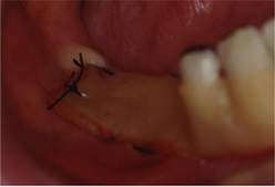
| Fig. 14. Protection with surgical plaster. |
|||||||||||||||||
| Implantology | |||||||||||||||||
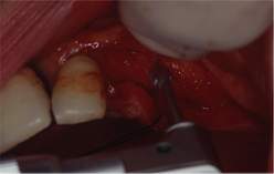 |
Fig. 15.
Surgical preparation of the bone that is to receive the implants in 2.4 and 2.5 and the periodontal mucoperiosteal access flap. |
||||||||||||||||
 |
 |
||||||||||||||||
|
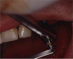
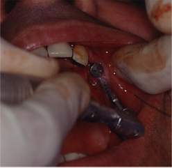
Figs 16, 17
Having inserted the first implant in 2.4, that of 2.5 is prepared. |
|||||||||||||||||
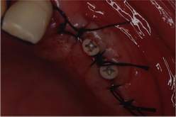 |
Fig. 18.
The two implants are inserted, the protective caps have been positioned and suturing of the two flaps has been completed with single stitches. |
||||||||||||||||
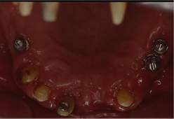 |
Fig. 19
The upper arch following periodontal and implantological surgery.The stumps are positioned on the three implants of 2.4 and 2.5 and of 1.4. Having maintained 2nd class mobility, but not presenting a periodontal pocket and showing an acceptable gain in bone, 2.1 was sacrificed as being unreliable and also in view of the complexity of the planned prosthesis, especially the upper denture with a horizontally pivoted shock-absorbing hinge. |
||||||||||||||||
 |
 |
||||||||||||||||
|
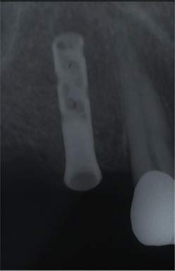
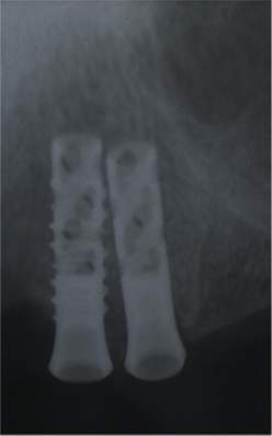
Figs 20, 21.
X-ray of implant in 1.4 and 2.4 and 2.5 respectively. |
|||||||||||||||||