|
|
|
|
|
|
|
|
|
|
|
|
|
|
|
|
|
| Lateral repositioning
of the flap |
 |
|
|
 |
|
|
|
|
| 15.
Above: the flap is shifted mesially: the dashed line indicates the
separation between the mucous flap (distal) and the mucoperiosteal
flap (mesial). |
|
| 14. Upper
left: the laterally positioned flap. After eliminating any periodontal
pockets that may be present or the epithelial sulcus, a mucous flap
is sculpted at the distal region and a mucoperiosteal flap is sculpted
at the mesial region. |
|
|
|
|
|
|
|
|
|
|
|
|
|
|
|
|
|
|
|
|
|
|
|
|
|
|
|
|
|
|
|
|
 |
 |
|
|
|
|
|
|
|
|
|
|
| 16. The flap
is positioned to cover the recession: distally (in red) we see the bed
of the donor site covered by the periosteum (in red in the figure to the
side). |
|
|
|
|
|
|
|
17. Recession on the mesial
root of 4.6. |
|
|
|
|
|
|
|
|
|
|
|
 |
 |
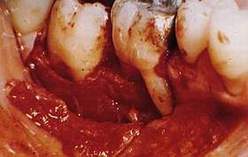
18. "V"-shaped
incision to eliminate the pocket corresponding to the recession.
|
19. A mucous flap is detached
at the distal region and a mucoperiosteal flap is is detached at the mesial
region. |
|
|
|
|
|
|
|
|
|
|
|
 |
 |
| 20. The flap
is sutured with single stitches. |
21. With the part of the
tissue remaining from the "design" of the flap a free gingival
graft is taken.21. With the part of the tissue remaining from the "design"
of the flap a free gingival graft is taken. |
|
|
|
|
|
|
|
|
|
|
|
 |
 |
|
|
|
|
|
|
|
|
|
|
|
|
|
|
|
|
| 22. The graft is sutured
onto the flap donor bed. |
|
|
|
|
|
|
|
|
|
23. In the picture on the
left we see an enlargement of the result of the operation: the recession
is covered and the adhering gum has increased.
|
|
|
|
|
|
|
|
|
|
|
|
|
|
|
|
|
|
|
|
|
|